

erythrocytesIDB (Version 2,
October 2017)
Goals
The creation of this database, which we call erythrocytesIDB, will advance the Erythrocytes shape classification. The motivation behind this dataset is that, as a research community, we need to compare a variety of different methods to find which choices are most important for a practical and robust solution.
Data description
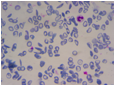
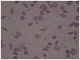
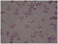
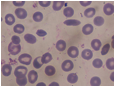
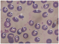
The erythrocytesIDB contains images of peripheral blood smears samples taken from patients with Sickle Cell Disease in Special Hematology Department of the General Hospital ‘Dr. Juan Bruno Zayas Alfonso’ from Santiago de Cuba. They are distributed like this:
erythrocytesIDB1
|
Description |
Example |
|
196 full field images |
|
|
629 images of individual cells classified as circular, elongated or other |
|

 e0001.jpg (elongated)
 o0001.jpg (other)
erythrocytesIDB2
|
Description |
Example |
|
50 full field images (source) |
01erythrocytesIDB2/source.jpg |
|
A labeled image, for each source image. |
01erythrocytesIDB2/labeled.jpg |
|
A mask image, for each source image. Binary image, cells in white. |
01erythrocytesIDB2/mask.jpg |
|
A mask image for circular cells, for each source image. Binary image, circular cells in white. |
01erythrocytesIDB2/mask-circular.jpg |
|
A mask image for elongated cells, for each source image. Binary image, elongated cells in white. |
01erythrocytesIDB2/mask-elongated.jpg |
|
A mask image for other cells, for each source image. Binary image, other cells in white. |
01erythrocytesIDB2/mask-other.jpg |


erythrocytesIDB3
|
Description |
Example |
|
30 full field images (source) |
01erythrocytesIDB3/source.jpg |
|
A labeled image, for each source image. |
01erythrocytesIDB3/labeled.jpg |
|
A mask image, for each source image. Binary image, cells in white. |
01erythrocytesIDB3/mask.jpg |
|
A mask image for circular cells, for each source image. Binary image, circular cells in white. |
01erythrocytesIDB3/mask-circular.jpg |
|
A mask image for elongated cells, for each source image. Binary image, elongated cells in white. |
01erythrocytesIDB3/mask-elongated.jpg |
|
A mask image for other cells, for each source image. Binary image, other cells in white. |
17erythrocytesIDB3/mask-other.jpg |


Adquisition
Images were obtained as follows:
A. Sample
preparation:
Samples were obtained from volunteer patients, using a lancet to obtain a drop of blood in a plate, that it was extended and dried, it was fixed with absolute alcohol and it was stained with Giemsa in a proportion of 2 % of reagent for 1 ml of distilled water. They were dried from 15 to 20 minutes again and washing with distilled water.
B. Image
acquisition
Images were taken with a Leika microscope with 100x augmented lens and a Kodak EasyShare V803 camera with Kodak Retinar Aspheric All Glass Lens of 36-108 mm AF 3X Optical lens, no professional.
C. Cell classification:
A 1st Grade specialist in Clinical Laboratory from the Special Hematology Department analyzed the images. Cells were classified in circular; elongated or cell with other deformations, and the specialist also determined the clusters with cells overlapped and the types of cells in those clusters.
People
· Pedro Marrero Fernández. Facultad de Matemática y Computación. Universidad de Oriente. Cuba.
· Grethel Coello Said. Facultad de Matemática y Computación. Universidad de Oriente. Cuba.
· Wilkie Ernesto Delgado Font. Facultad de Matemática y Computación. Universidad de Oriente. Cuba.
· Silena Herold Garcia. Facultad de Matemática y Computación. Universidad de Oriente. Cuba.
· Karelis Fernández Garcia. Hospital General “Dr. Juan Bruno Zayas Alfonso”. Cuba.
· Arquímides Montoya Padrón. Hospital General “Dr. Juan Bruno Zayas Alfonso”. Cuba.
· Manuel González-Hidalgo. Departament de Ciències Matemàtiques i Informàtica. Universitat de les Illes Balears. Spain
· Antoni Jaume-i-Capó. (erythrocytesIDB Coordinator) Departament de Ciències Matemàtiques i Informàtica. Universitat de les Illes Balears. Spain
DOWNLOAD
To obtain access erythrocytesIDB fill up next form:
erythrocytesIDB Release Agreement

